Trauma Physiology Laboratory
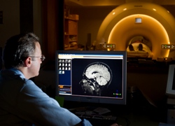
 Traumatic brain injury (TBI) affects approximately 1.7 million patients every year, but there are few options for medical management. We are using state-of-the art experimental techniques in calcium signaling, endothelial function, nitric oxide, and ion channels, to elucidate the molecular mechanisms and functional consequences of vascular responses to traumatic brain injury. Our clinical investigations using structural and functional MRI to study head injured patients are also providing new insight into the nature and progression of disease, as we develop novel therapies to ultimately improve patient outcomes after concussion. This research is closely aligned with the University's Neuroscience, Behavior and Health (NBH) Initiative.

Vascular Biology Techniques
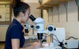
Equipment includes a video microscopy rig for imaging pressurized vessels, and a separate room with two dissection microscopes. We share the use of the nearby UVM Neurosurgery/ Skull Base Laboratory, founded by R.M.P. Donaghy, M.D., who invented many of the techniques of micro-neurosurgery in this same room, four decades ago. Key techniques used by our laboratory:
- Functional studies of pressurized arteries
- High-speed video calcium imaging of intact endothelium
- In vivo magnetic resonance imaging
 Nelson Laboratory
The Nelson laboratory is located in close proximity and houses the high-speed video laser microscopes used for calcium imaging. Dr. Mark Nelson, University Distinguished Professor and Chair of Pharmacology, is the mentor for Dr. Freeman’s NIH Clinical Scientist Research Career Development Award (K08) project, “Endothelial Ca2+ signals and vasodilatory function after traumatic brain injury”.